Medicine is a discipline defined by precision, complexity, and constant innovation. Yet much of its knowledge—whether at the molecular, cellular, or systemic level—remains inaccessible to the naked eye or difficult to explain through text alone. To bridge this gap, medical illustrations and animations have emerged as indispensable tools. They combine scientific accuracy with visual storytelling, turning intricate medical concepts into clear, engaging, and accessible visuals for learners, professionals, and patients.
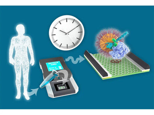
Table of Contents
The Power of Medical Illustrations
Medical illustrations https://scientific-illustrations.com/portfolio/medical-illustrations have a long tradition, dating back to early anatomical drawings that helped physicians understand the human body. Today, they are more advanced than ever, supported by digital technology and 3D rendering. What sets illustrations apart from photography is their ability to highlight the essential while excluding unnecessary details.
For example, a medical illustration of the respiratory system can focus on airflow through the lungs without distractions from surrounding tissues. This selective clarity makes illustrations invaluable for teaching anatomy, guiding surgical procedures, and supporting clinical research.
Medical illustrations serve multiple audiences. For students, they simplify complex material and provide visual anchors for memory. For healthcare professionals, they function as accurate reference tools during diagnosis or surgery. For patients, they explain conditions and treatments in a way that fosters understanding and trust.
Medical Animations: Bringing Biology to Life
While illustrations capture a single moment, medical animations https://scientific-illustrations.com/portfolio/animation bring processes into motion. They are especially effective in showing dynamic systems such as blood circulation, synaptic transmission, or the mechanism of action of new drugs. Animations allow viewers to observe sequences in real time, making invisible phenomena both visible and understandable.
In medical education, animations are used to simulate surgeries, demonstrate physiological processes, and explain pathologies. They enhance engagement and improve knowledge retention compared to text-only resources. In patient communication, animations can explain treatment steps, reduce anxiety, and encourage informed decision-making.
The pharmaceutical and biotechnology industries also rely on animations to demonstrate how therapies work at the cellular or molecular level, helping bridge the gap between research and application.
Applications Across Healthcare

Medical illustrations and animations now permeate every level of healthcare:
- Education: Medical schools integrate them into curricula to teach anatomy, physiology, and pathology more effectively.
- Training: Animations and illustrations help medical residents and professionals prepare for complex surgical procedures.
- Patient Care: Simplified visuals explain conditions and treatments, improving compliance and reducing uncertainty.
- Research Communication: Visual tools clarify findings, making research more accessible within the scientific community and beyond.
- Public Health: Campaigns use illustrations and animations to explain prevention strategies, encourage vaccination, and build public awareness.
Here’s a draft blog post based on your requirements. I chose the keyword: technology in medical imaging.
Meta Description:
Technology in medical imaging has reshaped healthcare. From X-rays to AI-driven 3D tools, learn how modern imaging improves diagnosis, surgery planning, and patient care.
How Technology Has Impact on Medical Illustrations
A single picture can sometimes reveal what words cannot. In healthcare, that picture often comes from medical imaging. From early X-rays to today’s AI-driven 3D models, technology in medical imaging has reshaped how doctors see inside the human body without making an incision.
As a medical imaging specialist, I’ve seen this progress firsthand. Years ago, I worked on a surgery helper software built using the VTK library. My goal was to create 3D models of a patient’s anatomy that could guide surgeons before they stepped into the operating room. That experience showed me how powerful medical imaging has become—not just for diagnosis but also for planning and improving surgical outcomes.
This article takes you through the journey of medical imaging: where it started, how it grew, and the role modern tools now play in healthcare.
A Brief History of Medical Illustrations
The Early Days
Medical imaging began in 1895, when Wilhelm Roentgen discovered X-rays. Suddenly, doctors could see bones inside the body without surgery. This changed how fractures were treated and gave medicine a new way of looking at hidden injuries.
Growth Through the 20th Century
- CT Scans (1970s): Created layered images of organs and tissues.
- MRI (1980s): Offered a clearer view of soft tissues without radiation.
- Ultrasound: Grew into a common tool for pregnancy monitoring and organ scans.
Digital Shift
As computers became stronger, imaging shifted from film to digital storage. This step allowed radiologists to share scans quickly, store them safely, and compare results across time.
How Technology Shapes Modern Medical Imaging
Here is some behind the scenes video of Medical Imaging from Mayo Clinic.
1. Better Visualization
Modern software turns flat scans into 3D models. For example, using the VTK library, I built a system that could generate accurate 3D structures of patient anatomy. Surgeons could rotate, zoom, and measure before operating, reducing surprises in the operating room.
2. Faster and Sharper Images
New machines capture images in seconds with better resolution. This reduces patient discomfort and helps doctors diagnose conditions early.
3. AI in Imaging
Artificial intelligence now helps radiologists spot patterns. AI tools can detect tumors, flag anomalies, and even suggest possible diagnoses, making the work faster and sometimes more accurate.
4. Minimally Invasive Surgery Support
By combining imaging with surgical navigation systems, doctors can operate with smaller cuts, less pain, and quicker recovery for patients.
Comparing Imaging Technologies
Here’s a quick look at how different imaging technologies serve various needs:
| Imaging Method | Best Use Case | Strengths | Limitations |
|---|---|---|---|
| X-ray | Bone fractures, lung scans | Fast, cheap | Limited detail of soft tissue |
| CT Scan | Brain, chest, abdomen | Detailed cross-sections | Radiation exposure |
| MRI | Brain, muscles, joints | Excellent soft tissue detail | Expensive, time-consuming |
| Ultrasound | Pregnancy, organ scans | Safe, no radiation | Image quality varies |
| PET Scan | Cancer, heart disease | Shows function & metabolism | Expensive, less available |
Personal Experience: Using Technology for Surgery Planning
When I built the surgery helper tool with VTK, my aim was to give surgeons more than a static image. By generating 3D models, they could plan cuts, estimate risks, and visualize blood vessels in relation to tumors.
During tests, surgeons shared that such tools saved time and reduced uncertainty. For complex surgeries, even small improvements in planning can mean safer outcomes. That project showed me that imaging isn’t just about looking inside—it’s about preparing for what comes next.
Impact on Surgery Planning and Outcomes
Safer Surgeries
Imaging gives surgeons a roadmap. Instead of going in blind, they know where arteries, nerves, and organs are. This reduces the risk of complications.
Shorter Operations
Clear planning cuts down operation time. That means less anesthesia and faster recovery for patients.
Better Patient Communication
3D models help doctors explain conditions to patients. Seeing a tumor in relation to nearby tissue can make treatment options easier to understand.
The Role of AI and Machine Learning
AI is no longer just a buzzword in healthcare—it’s already changing radiology.
- Early Cancer Detection: AI can highlight suspicious spots on mammograms.
- Brain Scans: Algorithms detect small strokes that a tired human eye may miss.
- Workflow Support: AI sorts through thousands of scans daily, flagging urgent cases first.
Still, AI doesn’t replace radiologists. It acts as an assistant, giving doctors more time to focus on complex decisions.
Telemedicine and Cloud Imaging
Technology in medical imaging is also linked to telemedicine. Cloud-based platforms allow doctors to access scans from anywhere. A radiologist in one country can read images taken in another. This improves access for rural areas where specialists may not be available.
Challenges in Medical Imaging
Even with progress, hurdles remain:
- High Costs: Advanced machines and software are expensive.
- Data Security: Cloud storage raises questions about patient privacy.
- Over-reliance on Technology: Doctors still need to confirm findings through clinical judgment.
Future Directions
Medical imaging is heading toward:
- Real-time 3D imaging during surgery.
- Portable imaging devices for field use.
- Greater AI integration for faster triage.
- Personalized scans based on genetic profiles.
Summary Table: How Technology Changed Medical Imaging
| Era | Technology | Impact |
|---|---|---|
| 1895 | X-rays | First internal view of the body |
| 1970s | CT scans | Detailed cross-sections |
| 1980s | MRI | Clearer soft tissue scans |
| 2000s | Digital Imaging | Faster sharing and storage |
| 2010s+ | AI & 3D modeling | Better planning and diagnosis |
Innovations in Medical Visualization
Advancing technologies are expanding the role of visuals in healthcare. Virtual reality (VR) and augmented reality (AR) offer immersive environments for training and patient education. A student can virtually explore human anatomy in three dimensions, while a surgeon can rehearse a procedure in a simulated operating room.
Artificial intelligence is beginning to assist in generating anatomical models and speeding up the production of complex medical visuals. Interactive platforms now allow learners and patients to manipulate visuals directly, turning passive observation into active exploration.
Another emerging trend is cinematic medical visualization, which merges the techniques of film and storytelling with scientific accuracy. This approach enhances not only clarity but also emotional impact, helping audiences connect with medical knowledge on a deeper level.
The Human Dimension of Visual Medicine
At the intersection of medicine and art lies collaboration. Scientists and healthcare professionals ensure accuracy, while illustrators and animators bring design skills and narrative power. Together, they create visuals that are precise, informative, and approachable.
These tools go beyond education and research—they humanize medicine. For patients, a clear illustration or animation can transform fear into understanding. For professionals, they provide confidence and clarity. For society at large, they ensure that vital medical knowledge is communicated effectively.
Conclusion
Technology in medical imaging has reshaped healthcare from the first X-ray to AI-powered analysis. Each step improved how doctors diagnose, plan, and treat patients. From my personal experience developing a VTK-based surgery planning tool, I’ve seen how 3D models can support surgeons in making safer decisions.
As medical imaging keeps growing, the goal stays the same: helping patients live longer, healthier lives. For those curious about where this field is going, watch the rise of AI-assisted tools, portable devices, and real-time surgical imaging. They will define the next stage of healthcare.
Medical illustrations and animations are more than visual aids—they are central to modern healthcare communication. They transform the invisible into the visible, simplify the complex, and connect science with human experience. As technology continues to evolve, their impact will only deepen, reshaping how medicine is taught, practiced, and shared. By merging accuracy with creativity, they ensure that the art of visualization remains at the heart of medical progress.